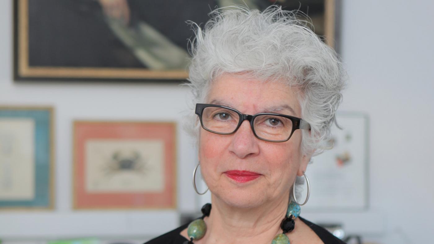
When Carol Mason, PhD, began studying the developing visual system nearly 30 years ago, scientists understood that our eyes are connected to our brain. But little was known about how the nerve cells making that connection form in the immature brain. Her research has helped to reveal the processes guiding their growth — opening up the possibility of repairing damage to the visual system caused by injury or disease.
Today the António Champalimaud Foundation celebrates the pioneering work of Dr. Mason and three other scientists who have investigated how visual signals move from the retina (a light-sensitive part of the eye) to the brain. The researchers share the Champalimaud Vision Award and a prize of one million euro (about $1.1 million) intended to further their investigations. The prize was announced at a ceremony in Lisbon, Portugal.
This is an exciting time to be studying how the brain is wired. We’re only just at the beginning.
“This is an incredible honor that will help to push our work in new directions,” said Dr. Mason, a neuroscientist, principal investigator at Columbia’s Mortimer B. Zuckerman Mind Brain Behavior Institute and professor of pathology and cell biology, neuroscience and ophthalmic science at Columbia University Medical Center. “This kind of open-ended funding gives you the freedom to do experiments outside of the box, take intellectual risks and go after big questions.”
For Dr. Mason, understanding the circuit that links the eyes to the brain and allows us to see is one of the most interesting puzzles in brain science. Sight begins with light striking cells, called photoreceptors, in the retina; those light detectors generate electrical signals picked up by cells in the eye, the retinal ganglion cells. Extensions from the retinal ganglion cells grow back through the skull to areas of the brain that process the electrical signals. The result is a composite image of the outside world.
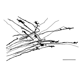
Above: Ink drawings of developing optic nerves changing direction as they grow from the eyes to the brain (Credit: Mason Lab/Columbia's Zuckerman Institute).
As they wind their way to the brain, the ganglion cell extensions, or fibers, take different paths and end up in different places. Some stay on the same side of the eye where they started; some cross over to the opposite side. This divergence of optic fibers helps to give us depth perception by sending information both to the left and the right sides of the brain.
But how does any growing cell in the embryo, which looks identical at first blush to its neighbors, know where to go?
“That’s the big question,” said Dr. Mason. "What endows cells that start at the same place, in the retina, with the ability to end up at different places in the brain?”
To find out, Dr. Mason and her colleagues began by staining growing cells with dyes. Looking at the cells under a microscope, she then meticulously traced their shapes by hand with pen and paper as they grew. Her drawings, photos and video movies early on showed the migrating tips of the ganglion cell axons, called growth cones, changing path in an x-shaped area of the head called the optic chiasm, just behind the eyes and below the brain.
Further work revealed molecules in this area that seemed to be serving as traffic cops, telling cells to go this way or that. Only growing nerves that carried a second molecule on the surface of their tips were directed to cross over to the other side of the brain.
Understanding what molecules influence growing nerve cells is a step toward regrowing damaged cells, said Dr. Mason. New therapies seek to program stem cells to repair injuries to the eyes and damage caused by diseases such as glaucoma or stroke.
“If you want to enable damaged ganglion cells to regenerate, you need to understand how to turn stem cells into retinal ganglion cells, with the right growth capabilities, and then guide them to the proper place to make their synaptic connections,” said Dr. Mason. “You need to understand the personalities of the cells and the factors that influence their development, and, we think, to recapitulate development.”
For Dr. Mason, the next frontier is understanding why different nerve cells ultimately connect not only to different sides of the brain but to different locales in the brain. She and others in the field have begun to compare the DNA of the different types of ganglion cells, looking for differences in what genes are turn on and off that could explain their ultimate fates.
“This is an exciting time to be studying how the brain is wired,” said Dr. Mason. “We’re only just at the beginning.”
Dr. Mason shares the prize with her colleagues Christine Holt, a professor at the University of Cambridge; John Flanagan, a professor of Harvard University; and Carla Shatz, a professor at the Stanford University School of Medicine.